WELCOME TO MY BLOG!

I am really happy you are here, because the people who read my blog are the reason I write and the motivation to keep doing this every day.

Thank you for being here and for wanting to find out more about me and my blog.

I sincerely appreciate you taking the time to visit my new blog. Please check back often.

Feel free to post comments and/or suggestions on any posts that you find of interest. Thanks a lot.....

Tuesday 24 July 2018

Respiratory Failure

Respiratory Failure

                                          Respiratory failure occurs when the respiratory system fails in either one or both of its gas exchange functions: oxygenation and the elimination of carbon dioxide. A reduction in the oxygen carried in the bloodstream is referred to as hypoxaemia, and a rise in the carbon dioxide levels within the bloodstream is referred to as hypercapnia.

Respiratory failure is classified into two groups


  •  Type I respiratory failure
  •  Type II respiratory failure


 Type I respiratory failure

                                                              Type I respiratory failure occurs when there is a problem with oxygenation resulting in hypoxaemia. This is most commonly caused by ventilation/perfusion mismatch resulting in reduced diffusion of oxygen from the alveoli into the pulmonary circulation. Type I respiratory failure is characterized by


Type II respiratory failure 

                                                              Type II respiratory failure occurs when there occurs when there is inadequate alveolar ventilation resulting in hypoxaemia and hypercapnia. Type II respiratory failure is characterized by


                                                                          Type II respiratory failure can be further sub-classified depending on the pre-existing condition of the patient and the speed of onset


Acute type II respiratory failure

                                                                        The patient will have no, or minor, evidence of pre-existing respiratory disease and patients typically have a high PaCO2, low pH, and normal bicarbonate.

Chronic type II respiratory failure

                                                                        Evidence of chronic respiratory disease, high PaCO2, normal pH, and high bicarbonate (>26 mmol/l).


Acute-on-chronic type II respiratory failure

                                                                            An acute deterioration in an individual with significant pre-existing type II respiratory failure, high PaCO2, low pH, and high bicarbonate (>26 mmol/l).

Myasthenia gravis (MG)


Mysthenia Gravis
Myasthenia gravis (MG) is either an autoimmune or congenital neuromuscular disease that leads to fluctuating muscle weakness and fatigue. The name myasthenia gravis, which is Latin and Greek in origin, literally means "grave muscle weakness." Myasthenia Gravis affects persons from all ethnic groups and both genders, although it commonly affects young adult women under the age of forty years, and men over the age of sixty. Myasthenia Gravis can affect persons of any age as well. On occasion, the disease can occur in members of the same family, although the disease is not contagious or directly inherited

Definition 
  • Chronic auto-immune disorder of the post-synaptic membrane at the neuromuscular junction (NMJ) in skeletal muscle
  • Circulating antibodies against the nicotinic acetylcholine receptor (AchR) and associated proteins impair neuromuscular transmission
  • Patients present with muscle weakness which typically worsens with continued activity (fatigue) and improves on rest
  • Severity varies from isolated eye muscle weakness to generalised muscle weakness and respiratory failure requiring mechanical ventilation
Risk Factors 
  • Female gender 
  • Ages 20 – 40
  • Familial myasthenia gravis
  • D-penicillamine ingestion (drug-induced myasthenia)
  • Having other autoimmune diseases
Differential diagnosis 
  • Lambert-Eaton myasthenic syndrome (LEMS)
    • Prominent proximal leg and arm weakness accompanied by diminished muscle stretch reflexes that improve with brief exercise.
    • Autonomic symptoms such as dry mouth, constipation and impotence are present in some patients.
    • Eye muscles are occasionally involved at presentation.
    • Respiratory failure is uncommon.
    • Associated with small-cell lung cancer in up to 70% of patients.
  • Botulism
    • Symptoms similar to myasthenia gravis (MG) but there may be hypotension, bradycardia, diarrhoea followed by constipation, and urinary retention. 
  • Penicillamine-induced myasthenia gravis 
    • Symptoms similar to MG can occur after weeks or months of treatment
    • They may remit on withdrawal of the drug; recovery may be slow or incomplete
  • Primary myopathies
    • Gradually progressive muscle weakness which is not fatigable.
    • These include auto-immune inflammatory myopathies or heritable myopathies as in mitochondrial diseases, such as progressive external ophthalmoplegia, or oculopharyngeal muscular dystrophy (OPMD).
    • Positive family history
Clinical features 
  • Muscle fatigability
    • The weakness worsens with activity (fatigue) and improves on rest and the fluctuations show diurnal variation (better in morning than in the evening)
  • Ptosis
    • Drooping eyelids and double vision occur early in the majority of patients.
    • The pupils are spared.
    • Cooling of eyelid for at least 2 minutes with an ice pack (ice test) improves ptosis in more than 95% of patients with myasthenia gravis (MG). 
      • It does not improve severe ptosis
  • Diplopia
    • Double vision occurs early in the majority of patients.
  • Dysphagia
    • Difficulties in chewing or swallowing occur when the facial and oropharyngeal muscles are affected
  • Dysarthria
    • Changes in speech occur when the oropharyngeal muscles are affected and there may be a characteristic nasal speech
  • Facial paresis
    • Changes in expression occur when the facial muscles are affected and there may be a characteristic flattened smile
  • Proximal limb weakness
    • Difficulty in getting out of chairs or climbing stairs. 
    • There is no evident muscle wasting. 
    • Reflexes are normal. 
    • Sensations are intact
  • Shortness of breath
    • If shortness of breath becomes severe enough to require mechanical ventilation, the patient is said to be in MG crisis
Pathophysiology 
  • In myasthenia gravis (MG), an auto-immune attack against acetylcholine receptors (AchRs) results in destruction of the post-synaptic membrane.
  • The reduced number of available binding sites for acetylcholine leads to inconsistent generation of muscle fibre action potentials which manifest as skeletal muscle weakness. 
  • Muscle tyrosine kinase (MuSK) is an agrin dependent protein on muscle membrane with an essential role in anchoring AchR at the tips of the postsynaptic folds
Investigations
  • Serum anti-acetylcholine receptor (anti-AchR) antibody analysis
    • Antibodies are detectable in 80% to 90% of patients with generalised myasthenia gravis (MG) and up to 50% of patients with ocular MG.
      • Sensitivity: greater than 90% in generalised MG; 40% to 60% in ocular MG. 
      • Specificity: 99%.
    • If the result is negative or equivocal proceed to antimuscle tyrosine kinase (MuSK) antibody assay
  • Antimuscle tyrosine kinase (MuSK) antibodies
    • Positive test in up to 70% of AchR seronegative generalised MG.
  • Serial pulmonary function tests
    • Indicated if shortness of breath and suspected MG crisis.
    • Serial measurements of FVC and negative inspiratory force (NIF) are taken. 
    • Indication for mechanical ventilation includes FVC 15 mL/kg or less (normal ≥60 mL/kg) and NIF 20 cm H2O or less (normal ≥70 cm H2O). 
    • Physicians should not wait for abnormal ABG as it occurs late in the course after clinical decompensation
  • Antistriational antibodies
    • This test is not done routinely in all patients
    • These antibodies are rare in patients without thymoma.
    • Detected in 75% to 95% of patients with thymoma and MG
  • Repetitive nerve stimulation
    • Sensitivity, 79% in generalised MG, 50% in ocular MG. 
    • Specificity, 97%.
    • Brief exercise of a muscle prior to the test may enhance decrement response.
    • If the result is negative and clinical suspicion is high, proceed to single fibre EMG.
    • Positive decrement also seen in Lambert-Eaton myasthenic syndrome (LEMS) and amyotrophic lateral sclerosis (ALS).
    • In MuSK MG, generally the yield of abnormal result is high in proximal muscles, for example the trapezius, deltoid and facial muscles. 
    • For patients with neck and respiratory weakness, it is important to evaluate clinically affected muscles as the test may be normal in limb and face muscles.
    • Greater than 10% decline in compound muscle action potential (CMAP) amplitude between the first and fourth potential in a train of 10 stimulations of the motor nerve at 2 to 3 Hz is considered a positive response
  • Single-fibre EMG
    • Sensitivity, 86% to 92% and specificity, 70% to 96% in facial muscles in ocular MG; 
    • Sensitivity and specificity, 98% in generalised MG.
    • Abnormal test may be seen in LEMS and ALS, inflammatory myopathies, or patients injected with botulinum toxin. 
    • Increase variability in motor latencies (jitter) or complete failure of neuromuscular transmission (block) in some muscle fibres
  • CT of chest
    • Should be performed in all newly diagnosed patients to detect thymoma (which occurs in about 15% of patients with MG) or thymic hyperplasia (which occurs in 75% of MG patients)
Management

a) conservative
  • Supportive care
    • Includes DVT prophylaxis; ulcer prophylaxis; adequate nutrition and hydration; and avoidance of infections and drugs that may worsen myasthenia symptoms.
b) medical 
  • Plasma exchange or intravenous immunoglobulin
    • Acute therapy for the recovery of transmission across neuromuscular junction
    • Plasma exchange has rapid response with onset usually after 2 to 3 sessions. 
      • Effects are temporary, lasting weeks. 
      • Therapy is expensive and requires hospitalisation.
    • Intravenous immunoglobulin (IVIG) is easy to administer but expensive
      • When patients respond, the onset is rapid within 4 to 5 days with maximal response apparent within 1 to 2 weeks
    • In patients with muscle tyrosine kinase (MuSK) MG, plasma exchange and immunoglobulin are used during acute exacerbations and crises. 
    • Observational studies indicate plasma exchange to be more effective than immunoglobulin.
  • Pyridostigmine
    • Patients with frequent symptoms should be treated with a cholinesterase inhibitor.
    • In patients with muscle tyrosine kinase (MuSK) MG, clinical response to cholinesterase inhibitor therapy is generally less favourable
      • Frequent muscarinic and nicotinic side effects, and sometimes even worsening of symptoms. 
      • Despite this poor response, most patients receive pyridostigmine due to its low side effect profile and low cost
  • Corticosteroids
    • Corticosteroids are used in older men with ocular myasthenia gravis (MG) and those with mild disease who fail pyridostigmine monotherapy
  • Immunosuppressant
    • Several options are available and include low-dose corticosteroids, azathioprine, mycophenolate mofetil, ciclosporin and tacrolimus
    • Patients with generalised myasthenia gravis (MG) with moderate symptoms usually require chronic corticosteroid maintenance therapy.
    • It is prudent to begin therapy with low dose and gradually titrate up towards maximum dose 
      • High doses can induce worsening of MG symptoms, including precipitating a myasthenic crisis.
    • Immunotherapy is tailored for each patient, including time to onset of response, adverse effect profile, availability and cost
    • Tacrolimus is generally better tolerated than ciclosporin.
c) surgical
  • Intubation and mechanical ventilation
    • Indication for mechanical ventilation includes FVC 15 mL/kg or less (normal ≥60 mL/kg) and negative inspiratory force (NIF) 20 cm H2O or less (normal ≥70 cm H2O)
  • Some patients may require a tracheostomy and percutaneous endoscopic gastrostomy (PEG) tube.
  • Thymectomy
    • Performed in patients with thymoma (any age) or without thymoma (15 to 55 years old). 
    • Approaches available include robotic thoracotomy, transcervical or full sternotomy. 
    • It is generally accepted that more complete the removal of thymus, higher is the rate of remission in MG symptoms.
    • Onset of benefit is delayed, rarely seen within 6 months and requires follow-up of up to 2 to 5 years for demonstrated efficacy.
    • The role of thymectomy for patients with muscle tyrosine kinase (MuSK) MG remains uncertain in the absence of a controlled trial.
Prognosis
  • Symptomatic improvement and clinical remission are the goals of therapy. 
    • However, the onset of improvement varies greatly from days to months. 
  • Typically, older men with ocular complaints respond promptly to corticosteroid monotherapy whereas generalised symptoms are slower to respond and require more aggressive therapy. 
  • Chronic maintenance drug therapy is often required. 
  • Disease exacerbations can occur due to infections, surgery, medicine exposures, malignancy, pregnancy or other stressors. 
    • Myasthenic crisis can sometimes be averted with aggressive early intervention during an exacerbation.
  • Most patients enjoy good quality of life and normal lifespan due to advances in diagnosis and immunosuppressive treatment.

Monday 23 July 2018

Apgar Score


Apgar score


The Apgar score is a simple assessment of how a baby is doing at birth, which helps determine whether your newborn is ready to meet the world without additional medical assistance. Your practitioner will do this quick evaluation one minute and five minutes after your baby is born.
The 1-minute score determines how well the baby tolerated the birthing process. The 5-minute score tells the health care provider how well the baby is doing outside the mother's womb.
In rare cases, the test will be done 10 minutes after birth.
This score – developed in 1952 by anesthesiologist Virginia Apgar and now used in modern hospitals worldwide.

What Does the Apgar Score Mean?

Apgar is an acronym, standing for the following criteria:
  • Appearance
  • Pulse (heart rate)
  • Grimace (reflexes)
  • Activity (muscle tone)
  • Respiration (breathing effort)
A pediatrician, ob-gyn, midwife or nurse will assign your newborn an Apgar score from 0 to 2 on each of the five criteria, with a total of 10 possible points. The higher the Apgar score, the better your baby is doing.
Appearance / (Skin color)
  • If the skin color is pale blue, the infant scores 0 for color.
  • If the body is pink and the extremities are blue, the infant scores 1 for color.
  • If the entire body is pink, the infant scores 2 for color.
Pulse / (Heart Rate)
Heart rate is evaluated by stethoscope. This is the most important assessment:
  • If there is no heartbeat, the infant scores 0 for heart rate.
  • If heart rate is less than 100 beats per minute, the infant scores 1 for heart rate.
  • If heart rate is greater than 100 beats per minute, the infant scores 2 for heart rate.
Grimace (reflexes)
Grimace response or reflex irritability is a term describing response to stimulation, such as a mild pinch:
  • If there is no reaction, the infant scores 0 for reflex irritability.
  • If there is grimacing, the infant scores 1 for reflex irritability.
  • If there is grimacing and a cough, sneeze, or vigorous cry, the infant scores 2 for reflex irritability.
Activity (muscle tone)

  • If muscles are loose and floppy, the infant scores 0 for muscle tone.
  • If there is some muscle tone, the infant scores 1.
  • If there is active motion, the infant scores 2 for muscle tone.
Respiration (breathing effort)
  • If the infant is not breathing, the respiratory score is 0.
  • If the respirations are slow or irregular, the infant scores 1 for respiratory effort.
  • If the infant cries well, the respiratory score is 2.


Well Newborn Care


Well Newborn Care

Delivery Room Care

* Place the infant skin to skin with the mother, once he is stable.
* Assess the Apgar score, at 1 and 5 min after birth.
* Perform a brief physical examination to check that the infant is
   healthy (has no major anomalies or birth injuries, his/her tongue &
   body appear pink, and has normal breathing).
* Examine the hips to rule out dislocation.
* Umbilical cord care.
         ► Fix the cord clamp 3-5 cm away from the umbilicus, and cut the
                cord using a scalpel.
         ► Examine for any abnormality (single umbilical artery).
         ► Wipe the umbilical stump with ethyl alcohol 70%.
* Identification: take footprints and record in the medical record,
   and place 2 bracelets with identical hospital numbers (one on the
    wrist and the other on the ankle).

Transitional Care (first 4-6 hrs after birth)

* Common signs of disordered transitioning
         ► Respiratory distress
         ► Poor perfusion with cyanosis or pallor
         ► Need for supplemental oxygen
         ► Hypothermia
* Evaluate every 30-60 min
         ► Assess HR, RR, axillary temperature, color & tone.
* With suspicion of disordered transitioning
         ► If stable → observe closely for a period of time.
         ► If persistent signs → transfer to a higher level of care.

Routine Care

* Keep newborn with mother all the time (rooming-in).
* Perform proper hand washing before handling the newborn.
* Maintain newborn’s temperature
        ► Encourage skin to skin contact with the mother
        ► Use hats and proper clothes
* Assess GA using the expanded Ballard Score. Measure and
   record the newborn's weight, head circumference, and length,
   and then plot against the estimated GA.
* Bathing
        ► Do not bathe immediately after birth; vernix caseosa does not
             need to be removed.
        ► The first bath can be given with non-medicated soap and
             warm tap water once infant's temperature has stabilized (4-6
             hrs after delivery).
        ► Do not bathe the infant in a basin until after the umbilical
             stump has fallen off.
* Examine skin for trauma or signs of infection.
* Umbilical cord care
        ► Keep the cord dry and loosely covered with clean sterile gauze.
        ► Fold the diaper below the umbilicus.
        ► If soiled, wash with soap and clean water and dry it well.
        ► Apply alcohol after each diaper change.
* Place the newborn infant supine (on the back) to sleep and not
   prone (on the stomach).
* Routine medications
         ► Give vitamin K1 (0.5-1 mg IM) within 2 hrs of life.
* Feedings
         ► Support immediate and exclusive breastfeeding during the
              first hr postpartum preferably in the Delivery Room.
         ► Offer standard term formula to infants for whom breastfeeding
              is contraindicated at least every 3-4 hrs.

Instructions to the Mothers or Other Care-Givers

* Observe baby’s temperature, respiration & effort at feeding.
* Observe for passage of urine and stools.

Vaccination

* Educate parents about vaccination schedule.
* Administer HBIG (0.5 ml/kg IM) to all newborns of HBsAg positive
   mothers as soon as possible after birth (within 12 hrs),
   followed by HBV vaccine (0.5 ml IM).

Caring for life...


Can you wake up a Section officer at mid night to sign a document?

Can you wake up a clerk at mid night to prepare a bill?

Can you wake up a lecturer at mid night to teach you?

Can you wake up a shopkeeper to buy a match box from his shop?

Everybody will answer No

But you can wake up a NURSE from a deep sleep at mid night for a patient who suffering in pain,
We the Nurse stand at the bedside of patients to save them because we feel the pain inside us.

                          PROUD 2 B A NURSE


Sunday 22 July 2018

Nurses are the Real Life Heroes


          Critical-care nursing, or intensive-care-unit (ICU) nursing, is a specialty that requires focus and stamina. ICU nurses need a solid foundation of experience to actively monitor and treat acutely ill patients with life-threatening conditions. Critical-care Nurses are the real life heroes, always ready to fight against Pathogenic organisms and of course also against Angel of Death even lots of stressful environment.   

Glaucoma


GLAUCOMA

Glaucoma is a leading cause of blindness and visual impairment. Glaucoma, known as the “sneak thief of sight,” can affect patients of all ages. Many people affected with glaucoma do not experience any symptoms and may not be aware that they have the disease until they have lost a significant amount of vision.
 With early detection and treatment, eyes can be protected against the serious loss of vision or blindness. Glaucoma can affect anyone from newborn infants to the elderly.
TYPES OF GLAUCOMA

There are several types of glaucoma with two main types: 
open-angle and angle-closure.

OPEN-ANGLE GLAUCOMA
Glaucoma Treatment | Lasik Surgery | Ardmore OK | Lone Grove OK
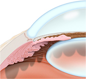
Open-angle glaucoma is the most common type of glaucoma where the fluid in the eye drains too slowly through the network of tiny drainage channels, known as the trabecula. The pressure in the eye increases as the fluid in the eye continues to build. Loss of vision occurs gradually and the vision loss is not always noticed until it becomes irreversible. About 95 percent of glaucoma cases are due to open-angle glaucoma.
ANGLE-CLOSURE GLAUCOMA
Glaucoma Treatment | Lasik Surgery | Ardmore OK | Lone Grove OK
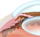
Angle-closure glaucoma occurs when the tiny drainage channels, known as the trabecula, become blocked which then causes a sudden rise in pressure in the eye. This condition is not common but when it occurs it requires immediate medical attention.

RISK FACTORS FOR GLAUCOMA

Everyone is at risk for developing glaucoma. Those at greater risk meet the following criteria:
  • Are at least 45 years old without regular eye exams
  • Have a family history of glaucoma
  • Have abnormally high eye pressure
  • Are of African descent
  • Are Hispanic and older than 60
  • Are nearsighted
  • Have thin corneas
  • Have diabetes
  • Have a previous eye injury
  • Regularly use cortisone/steroid products

DIAGNOSIS OF GLAUCOMA

To detect glaucoma, the physician will perform the following tests:
  • Visual acuity
  • Visual field
  • Tonometry
  • Dilated-eye examination
  • Retinal evaluation
  • Pachymetry
  • Gonioscopy

TREATMENT OF GLAUCOMA

Once glaucoma has been diagnosed, treatment should begin as soon as possible to help minimize the risk of permanent vision loss. There is no cure for glaucoma, so treatment focuses on relieving symptoms and preventing further damage. Treatment for each individual case depends on the type and severity of the glaucoma. Some of the treatment methods for glaucoma are:
MEDICATION
Eye drops or oral medication may be used to either reduce fluid production in the front of the eye or to help drain excess fluid. Side effects of the medication may result in redness, stinging, irritation or blurry vision.
LASER SURGERY
Trabeculoplasty, iridotomy or cyclophotocoagulation are laser procedures that aim to increase the outflow of fluid from the eye or eliminate fluid blockages.
SURGERY
A trabeculectomy may be performed to create a new channel to drain fluid from the eye and reduce the pressure that causes glaucoma.

love pics